If you live in Oxford, cycling is difficult to avoid. But as anyone new to the city can attest, hopping back on a saddle for the first time in years to weave through the narrow busy streets can be a daunting prospect. Luckily, the old saying holds true: it really is like learning to ride a bike. Many people will have experienced this phenomenon before, the amazing and long-lasting memory for skills that is often known as muscle memory.
We received lots of questions as part of the Big Brain Competition about muscle memory in all kinds of different skills, from knitting, to dancing, to gaming. What is muscle memory? Does it involve changes in to brain structure? What is happening in the brain when we learn something new?
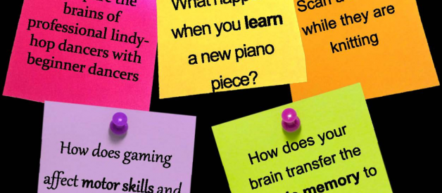
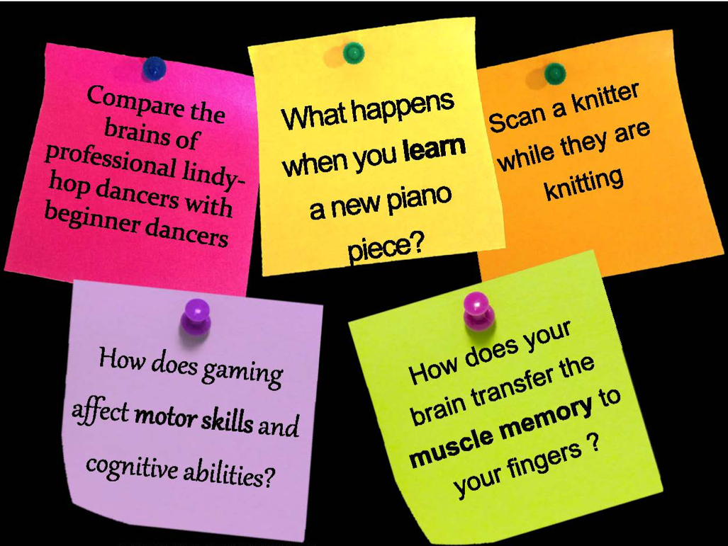 Questions submitted by members of the public as part of The Big Brain Competition.
Even the simplest everyday actions involve a complex sequence of tensing and relaxing many different muscles. For most of these actions we have had repeated practice over our lifetime, meaning that these actions can be performed faster, more smoothly and more accurately. Over time, with continual practice, actions as complicated as riding a bike, knitting, or even playing a tune on a musical instrument, can be performed almost automatically and without thought.
We often talk about these skills as being held in muscle memory, but this term is really a bit of a misnomer. Although certain skills, like cycling or perfecting a tennis serve, might require the strengthening of certain muscles, the processes that are important for learning and memory of new skills occur mainly in the brain, not in the muscles. Changes that occur in the brain during skill learning and memory alter the information that the brain sends out to the muscles, thereby changing the movements that are produced.
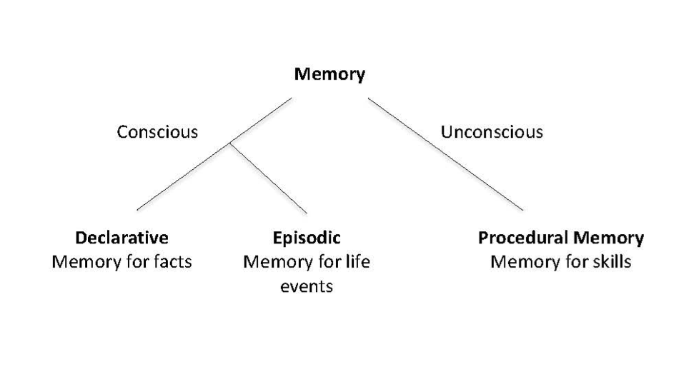
Despite this, skill learning and memory is clearly quite different from other forms of memory. It is thought that human memory is made up of multiple different systems that can all operate almost independently of each other[1]. For example we can have memories for facts, like the fact that Paris is the capital of France, but may not be able to remember when or where we originally learned this fact. Likewise, you might remember having a conversation with a friend, but not remember what the conversation was about. This is because the memory for facts, known as declarative memory, is thought to be a different system, controlled by different brain mechanisms, than the one used for memory of life events, known as episodic memory.
Memory for skills can be thought of as another distinct system. For example, you may be able to ride a bike perfectly, but that doesn?t mean you could explain to someone the exact sequence of movements needed in order to cycle. You may not even remember when or where you learned this skill. Experiments on patients with amnesia and other memory disorders have demonstrated how these different memory systems can operate separately. One patient, known as H.M., who suffered severe amnesia after surgery to cure epilepsy, and was unable to form new memories for life events or facts, had normal learning and memory for skills such as mirror drawing[2]. In this task H.M. would be asked to draw a simple image, like a star, while only seeing the image and his hand in a mirror, meaning his actions had to be made in the opposite direction to how they appeared to him. Amazingly, despite becoming highly skilled at mirror drawing, H.M. could never recognise the task equipment or remember any of the training sessions. This finding points out an important aspect of skill memory, that it can be stored without any conscious awareness, and the skilled actions can be performed almost automatically.
These different types of memory are controlled by different brain regions, with declarative and episodic memories mainly being produced and stored in the temporal lobe and hippocampus. Quite a large range of brain areas seem to be responsible for skill memories, including: areas in motor cortex, the part of the brain which sends signals to the muscle of the body and is responsible for planning and executing movements; the basal ganglia, a structure deep inside the brain which is associated with movement initiation; and the cerebellum, an area at the back of the brain which deals with adaptation.
But what happens to these regions when we learn something new? And what is it about these changes that allow improvement and memory for skills?
How does brain structure change when we make a skill memory?
Using magnetic resonance imaging (MRI), researchers can study the many different types of changes that allow us to learn and remember a motor skill. One of these changes involves increasing the connections between the different areas of the brain that are required for a particular skill. In one study, performed in Oxford, healthy adults had MRI scans before and after six weeks of juggling training. These scans could detect white matter, the long fibres that connect different parts of the brain together. The researchers found that after the juggling training there was an increase in the white matter connections between regions of the brain responsible for vision and regions responsible for making movements[3]. The increased connections between visual and movement areas results in faster and easier sharing of information, perhaps allowing for greater hand-eye coordination.
It is not just white matter that can change with training: studies have shown that there are changes in grey matter as well. Grey matter is made up of the brain cell (neuron) bodies, and is where information processing in the brain occurs. Another juggling study showed that after training there were increases in grey matter in parts of the brain that are involved in the processing of visual information about moving objects[4], perhaps allowing the visual information about the moving juggling balls to be processed more accurately.
Learning of new skills also results in changes in the primary motor cortex, the area of the brain responsible for causing actions. Cells in this area make connections with other neurons that travel down the spinal cord to contact the muscles of the body and cause them to contract. Parts of the body that are close to each other, such as the fingers, are controlled by areas that are close to each other in the motor cortex. We can safely and easily study how different parts of the motor cortex connect to muscles in healthy humans using a technique called transcranial magnetic stimulation (TMS). We use TMS to apply small magnetic pulses to the surface of the scalp in different places and record twitches in the muscles of the body.
Research using TMS and other techniques has discovered that ?representations? of the muscles of the body in the motor cortex vary between individuals depending on their use. For example, professional players of stringed instruments tend to have larger areas representing their left hand [5,6]. Having a larger representation, and so a greater number of connections from the brain to the muscles of the hand, perhaps allows for finer movement control. Although these changes are probably the result of years of intensive practice, small changes in representation can also occur over much shorter periods. One study asked healthy volunteers to learn a short hand and foot movement task. Results showed that the area of the motor cortex representing the hand muscles spread temporarily towards the area representing the foot[7]. Changing how the brain connects to the muscles is likely to be another way of improving skills, and if these changes are permanent then the skill will be preserved.
Changes in white matter, grey matter and in motor cortex representation all appear to be important for skill learning and memory. The brain of a person who is very good at a particular skill, such as lindy-hop dancing or playing a certain video game, might have stronger white matter connections between the different brain areas needed for each task, more grey matter in some of these regions, and might have larger motor cortex representations of the muscles needed. However, there are probably many other types of structural changes that occur when we learn a new motor skill that are yet to be discovered.
What about changes in brain function?
As well as measuring changes in brain structure, MRI scanners can also be used to look at brain function when performing different tasks. MRI can tell us about how brain activation changes as we learn new motor skills. Studies have shown that at the very beginning of learning a new movement there is a large amount of activity across the brain, but particularly in an area known as the pre-motor cortex, which lies just in front of the primary motor cortex[8], and is normally associated with movement planning. High levels of activity are also seen in the basal ganglia which is an area normally active during movement initiation [9,10]. The high levels of activity in these areas are probably related to the fact that, in order to learn a new skill, each action has to be planned and thought through. After repeated practice of the action, as it becomes an effortless almost automatic skill, the activity in the pre-motor cortex and basal ganglia decreases[11,12].
Other areas such as the motor cortex and the cerebellum remain active even when the action has become automatic, but activity here becomes more focussed[11]. These findings have been interpreted as the brain learning the most efficient way to perform the action. If we were to scan a knitter while they were learning a new stitch, or a gamer playing a new video game we would probably see roughly this pattern of activity.
What if we compared a novice knitter and a professional knitter while they leant this new stitch? Well, one study scanned both professional and amateur violinists while they performed the movements that would be required in order to play a section of a Mozart concerto. While there were many similarities in the activation, with both groups showing activation of the motor cortex, there was more focussed activation in the professional group, indicating their improved efficiency in preforming these movements. There were also fewer other brain areas, such as the basal ganglia, showing activation in the professional group, indicating that they performed the task more automatically than the amateur group[13].
So whether you?re a cyclist, a knitter, a dancer or a gamer you can thank similar changes in your brain structure and function for allowing you to improve and remember these skills. Without the amazing phenomenon of muscle or skill memory, none of this would be ?as easy as learning to ride a bike?.
Written by
Ainslie Johnstone, DPhil Student in the Wellcome Centre for Integrative Neuroimaging.
References
1. Squire, L. R. & Zola, S. M. Structure and function of declarative and nondeclarative memory systems. Proc. Natl. Acad. Sci. 93, 13515?13522 (1996).
2. Milner, B. Les troubles de la mmoire accompagnant des lsions hippocampiques bilatrales. Physiol. Hippocampe Cent. Natl. Rech. Sci. pp. 257?272 (1962).
3. Scholz, J., Klein, M. C., Behrens, T. E. & Johansen-Berg, H. Training induces changes in white matter architecture. Nat. Neurosci. 12, 1370?1371 (2009).
4. Draganski, B. et al. Changes in grey matter induced by training. Nature 427, 311 (2004).
5. Elbert, T., Pantev, C., Wienbruch, C., Rockstroh, B. & Taub, E. Increased Cortical Representation of the Fingers of the Left Hand in String Players. Science 270, 305 (1995).
6. Hashimoto, I. et al. Is there training-dependent reorganization of digit representations in area 3b of string players? Clin. Neurophysiol. 115, 435?447
7. Liepert, J., Terborg, C. & Weiller, C. Motor plasticity induced by synchronized thumb and foot movements. Exp. Brain Res. 125, 435?439 (1999).
8. Toni, I., Krams, M., Turner, R. & Passingham, R. E. The Time Course of Changes during Motor Sequence Learning: A Whole-Brain fMRI Study. NeuroImage 8, 50?61 (1998).
9. Seitz, R. J. & Roland, P. E. Learning of sequential finger movements in man: a combined kinematic and positron emission tomography (PET) study. Eur. J. Neurosci. 4, 154?165 (1992).
10. Seitz, R. J., Roland, P. E., Bohm, C., Greitz, T. & Stone-Elander, S. Motor learning in man: a positron emission tomographic study. Neuroreport 1, 57?60 (1990).
11. Wu, T., Kansaku, K. & Hallett, M. How Self-Initiated Memorized Movements Become Automatic: A Functional MRI Study. J. Neurophysiol. 91, 1690 (2004).
12. Karni, A. et al. Functional MRI evidence for adult motor cortex plasticity during motor skill learning. Nature 377, 155 (1995).
13. Lotze, M., Braun, C., Birbaumer, N., Anders, S. & Cohen, L. G. Motor learning elicited by voluntary drive. Brain 126, 866?872 (2003).
What next?
Follow us here on Medium where we?ll be publishing more articles soon.
If you liked this article please ?applaud? it to help spread the word and let others find it.
Want to read more? Try our articles on: How is your lifestyle affecting your brain?, Behind the scenes at the Brain Diaries Exhibition and Oxford and the mosquito.
