All animal cells are multicellular. They are eukaryotic cells. Animal cells are surrounded by a plasma membrane and it contains the nucleus and organelles that are membrane-bound. Unlike the eukaryotic cells of plants and fungi, animal cells do not have a cell wall. This feature was lost in the distant past by the single-celled organisms that gave rise to the kingdom Animalia.
Animal cells are of various sizes and have irregular shapes. Most of the cells size range between 1 and 100 micrometers and are visible only with the microscope. Trillions of cells are found in the human body. There are many different types of cells, approximately 210 distinct cell types in the adult human body.
The lack of a rigid cell wall allowed animals to develop a greater diversity of cell types, tissues, and organs. Specialized cells that formed nerves and muscles ? tissues impossible for plants to evolve ? gave these organisms mobility. The ability to move about by the use of specialized muscle tissues is a hallmark of the animal world, though a few animals, primarily sponges, do not possess differentiated tissues. Notably, protozoans locomote, but it is only via nonmuscular means, in effect, using cilia, flagella, and pseudopodia.
The animal kingdom is unique among eukaryotic organisms because most animal tissues are bound together in an extracellular matrix by a triple helix of protein known as collagen. Plant and fungal cells are bound together in tissues or aggregations by other molecules, such as pectin. The fact that no other organisms utilize collagen in this manner is one of the indications that all animals arose from a common unicellular ancestor. Bones, shells, spicules, and other hardened structures are formed when the collagen-containing extracellular matrix between animal cells becomes calcified.
Animals are a large and incredibly diverse group of organisms. Making up about three-quarters of the species on Earth, they run the gamut from corals and jellyfish to ants, whales, elephants, and, of course, humans. Being mobile has given animals, which are capable of sensing and responding to their environment, the flexibility to adopt many different modes of feeding, defense, and reproduction. Unlike plants, however, animals are unable to manufacture their own food, and therefore, are always directly or indirectly dependent on plant life.
Most animal cells are diploid, meaning that their chromosomes exist in homologous pairs. Different chromosomal ploidies are also, however, known to occasionally occur. The proliferation of animal cells occurs in a variety of ways. In instances of sexual reproduction, the cellular process of meiosis is first necessary so that haploid daughter cells, or gametes, can be produced. Two haploid cells then fuse to form a diploid zygote, which develops into a new organism as its cells divide and multiply.
The earliest fossil evidence of animals dates from the Vendian Period (650 to 544 million years ago), with coelenterate-type creatures that left traces of their soft bodies in shallow-water sediments. The first mass extinction ended that period, but during the Cambrian Period which followed, an explosion of new forms began the evolutionary radiation that produced most of the major groups, or phyla, known today. Vertebrates (animals with backbones) are not known to have occurred until the early Ordovician Period (505 to 438 million years ago).
Cells were discovered in 1665 by British scientist Robert Hooke who first observed them in his crude (by today?s standards) seventeenth century optical microscope. In fact, Hooke coined the term ?cell?, in a biological context, when he described the microscopic structure of cork like a tiny, bare room or monk?s cell. Illustrated in Figure 2 is a pair of fibroblast deer skin cells that have been labeled with fluorescent probes and photographed in the microscope to reveal their internal structure. The nuclei are stained with a red probe, while the Golgi apparatus and microfilament actin network are stained green and blue, respectively. The microscope has been a fundamental tool in the field of cell biology and is often used to observe living cells in culture. Use the links below to obtain more detailed information about the various components that are found in animal cells.
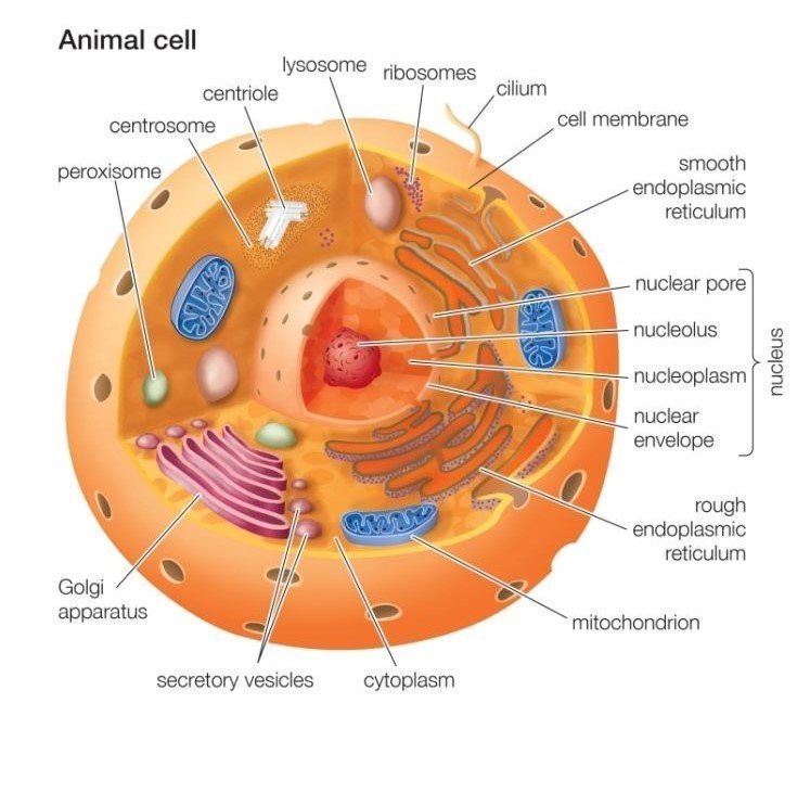
Cell membrane
It is a semi-permeable barrier, allowing only a few molecules to move across it. Electron microscopic studies of cell membrane show the lipid bilayer model of the plasma membrane, it is also known as the fluid mosaic model. The cell membrane is made up of phospholipids which has polar(hydrophilic) heads and non-polar (hydrophobic) tails.
Cytoplasm
The fluid matrix that fills the cell is the cytoplasm. The cellular organelles are suspended in this matrix of the cytoplasm. This matrix maintains the pressure of the cell, ensures the cell doesn?t shrink or burst.
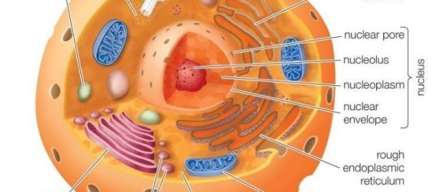
Nucleus
The nucleus is the house for most of the cells genetic material the DNA and RNA. The nucleus is surrounded by a porous membrane known as the nuclear membrane. The RNA moves in/out of the nucleus through these pores. Proteins needed by the nucleus enter through the nuclear pores. The RNA helps in protein synthesis through the transcription process. The nucleus controls the activity of the cell and is known as the control center. The nucleolus is the dark spot in the nucleus, and it is the location for ribosome formation.
Ribosomes
Ribosomes are the site for protein synthesis where the translation of the RNA takes place. As protein synthesis is very important to the cell, ribosomes are found in large number in all cells. Ribosomes are found freely suspended in the cytoplasm and also are attached to the endoplasmic reticulum.
Endoplasmic reticulum
ER is the transport system of the cell. It transports molecules that need certain changes and also molecules to their destination. ER is of two types, rough and smooth. ER bound to the ribosomes appear rough and is the rough endoplasmic reticulum; while the smooth ER does not have the ribosomes.
Lysosomes
It is the digestive system of the cell. They have digestive enzymes helps in breakdown the waste molecules and also help in detoxification of the cell. If the lysosomes were not membrane-bound the cell could not have used the destructive enzymes.
Centrosomes
It is located near the nucleus of the cell and is known as the ?microtubule-organizing center? of the cell. Microtubules are made in the centrosome. During mitosis the centrosome aids in dividing the cell and moving of the chromosome to the opposite sides of the cell.
Vacuoles
They are bound by a single membrane and small organelles. In many organisms, vacuoles are storage organelles. Vesicles are smaller vacuoles which function for transport in/out of the cell.
Golgi Bodies
Golgi bodies are the packaging center of the cell. The Golgi bodies modify the molecules from the rough ER by dividing them into smaller units with a membrane known as vesicles. They are flattened stacks of membrane-bound sacs.
Mitochondria
Mitochondria is the main energy source of the cell. They are called the powerhouse of the cell because energy(ATP) is created here. Mitochondria consists of the inner and outer membrane. It is spherical or rod-shaped organelle. It is an organelle which is independent as it has its own hereditary material.
Cilia and Flagella
Cilia and flagella are structurally identical structures. They are different based on the function they perform and their length. Cilia are short and are in large number per cell while flagella are longer and are fewer in number. They are organelles of movement. The flagellar motion is undulating and wave-like whereas the ciliary movement is power stroke and recovery stroke.
Endosomes and Endocytosis
Endosomes are membrane-bound vesicles, formed via a complex family of processes collectively known as endocytosis, and found in the cytoplasm of virtually every animal cell. The basic mechanism of endocytosis is the reverse of what occurs during exocytosis or cellular secretion. It involves the invagination (folding inward) of a cell?s plasma membrane to surround macromolecules or other matter diffusing through the extracellular fluid.
Intermediate Filaments
Intermediate filaments are a very broad class of fibrous proteins that play an important role as both structural and functional elements of the cytoskeleton. Ranging in size from 8 to 12 nanometers, intermediate filaments function as tension-bearing elements to help maintain cell shape and rigidity.
Microfilaments
Microfilaments are solid rods made of globular proteins called actin. These filaments are primarily structural in function and are an important component of the cytoskeleton.
Microtubules
These straight, hollow cylinders are found throughout the cytoplasm of all eukaryotic cells (prokaryotes don?t have them) and carry out a variety of functions, ranging from transport to structural support.
Peroxisomes
Microbodies are a diverse group of organelles that are found in the cytoplasm, roughly spherical and bound by a single membrane. There are several types of microbodies but peroxisomes are the most common.
In addition the optical and electron microscope, scientists are able to use a number of other techniques to probe the mysteries of the animal cell. Cells can be disassembled by chemical methods and their individual organelles and macromolecules isolated for study. The process of cell fractionation enables the scientist to prepare specific components, the mitochondria for example, in large quantities for investigations of their composition and functions. Using this approach, cell biologists have been able to assign various functions to specific locations within the cell. However, the era of fluorescent proteins has brought microscopy to the forefront of biology by enabling scientists to target living cells with highly localized probes for studies that don?t interfere with the delicate balance of life processes.

