?Well, this company uses EEG to tell me which areas of the brain are active when people watch my ad/hear my candidate/see my product/taste my soup?. I?m sorry to be the bearer of bad news? but they really don?t.
One of the most important aspects that buyers of Consumer Neuroscience must be aware of is the technical characteristics and limitations of the tools used by the vendors in their research projects. Spatial vs temporal resolution is a big one.
Classically, neuroimaging techniques have been classified by their spatial vs. temporal resolution (here?s a nice illustration at Grinvald and Hildesheim, 2004). In a nutshell, spatial resolution refers to the capacity a technique has to tell you exactly which area of the brain is active, while temporal resolution describes its ability to tell you exactly when the activation happened.
Usually, fMRI is described as the hallmark of spatial resolution: fMRI?s are now able to locate specific foci of activation with millimetre precision, although they are not that great in telling you when this activation happened.
On the other hand, EEG is the hallmark of temporal resolution ? because it is a measure of electrical activity that spreads virtually instantly, this technique can have millisecond resolution. What EEG is not that great at, however, is in telling you exactly where that activity was generated. This happens for a for a variety of reasons, the most important being that on the way from the neurons to the electrodes in the head, electrical current travels through a multitude of tissues (brain, cerebrospinal fluid, skull and scalp) with different conducting properties, flowing though the path of less resistance and being distorted along the way.
The estimation of the neural sources of EEG is, as von Helmholtz described back in 1853, an ill-posed problem, meaning many different source combinations can result in the same reading at the scalp. Although there are some nice algorithms (if you?re into it) of estimating the neural sources of EEG signals from the activity recorded in the scalp (this is called ?solving the inverse problem?), this requires extremely clean recordings (you definitely can?t be walking along a supermarket shelf wearing an EEG band) and a high number of recording electrodes (less than 32 is really bad, the quality improves dramatically until 68 and plateaus at around 100 electrodes ? Michel et al., 2004).
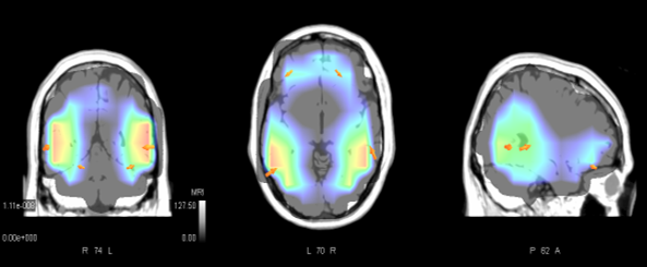 This source location, for a really clean EEG recording from 32 electrodes, estimates that the signal is basically located somewhere in the head!
The bottom line is: next time someone tells you that ?area X? of the brain is activated by your product using (most of the times low-density) electroencephalography, ask them to show you their new revolutionary EEG source estimation algorithm. If they have one, tell them they should publish it in a high-impact journal and help advance fields such as epilepsy monitoring (where these algorithms are mostly researched and really useful). If they don?t (which they most likely won?t) ? oh well?

